Watch
Overview
At Allevia Radiology, we provide leading services of nuclear medicine in Auckland, offering both diagnostic and therapeutic procedures using safe, low-dose radioactive tracers.
Allevia Radiology's Theranostics & Research Team offer leading nuclear procedures, providing an array of different treatments for a wide variety of conditions. Our team has experience in experimental and compassionate use of radiopharmaceuticals for therapy, and initiates and participates in local and international clinical trials regularly, ensuring we can offer the latest care and advances in nuclear medicine in New Zealand.
We offer consultations to patients with appropriate referrals from specialists or oncologists, discussing the variety of therapy options for oncological and non-oncological nuclear medicine therapy to help treat your specific condition.
What is Nuclear Medicine?
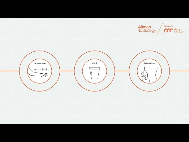
Nuclear medicine is the branch of medicine that involves the administration of radiopharmaceuticals in order to diagnose and treat disease. The scanner we use produces images by detecting a small amount of a radioactive tracer in your body, which is either injected, swallowed or inhaled.
The main difference between nuclear medicine and other imaging modalities is that nuclear imaging shows how the tissue or organ being scanned is functioning, while the traditional systems such as computed tomography (CT) and magnetic resonance imaging (MRI scan) show only the anatomy or structure.
SPECT-CT Scans
‘SPECT' imaging allows us to view nuclear medicine scans in 3D and on multiple different planes, which increases sensitivity and overall accuracy of diagnosis. The combination with “multi-slice CT” adds further diagnostic information by also allowing for very accurate anatomical localisation.
Our hybrid multi-slice SPECT-CT is one of the most advanced scanners in New Zealand.
SPECT-CT imaging can assist in diagnosing Alzheimer’s
Alzheimer’s disease is one of the most important neuro-degenerative conditions which usually presents with progressive memory loss. There is now general agreement that Alzheimer’s is amenable to diagnosis with SPECT-CT scanning.
In the early stages of disease, standard imaging with CT or MRI usually show normal findings, however, SPECT nuclear imaging of the brain can demonstrate abnormalities in the perfusion patterns of the brain (cerebral hemispheres) before the onset of structural changes.
What are the types of Nuclear Medicine?
We take pride in offering a fully integrated radiology reporting service. This means that all relevant information from other scans, e.g. plain radiographs, MRI, ultrasound and CT, is taken into account, alongside a wide variety of nuclear medicine scans to further assist in diagnosing and treating your condition.
Brain Perfusion
Brain Perfusion
Cerebral perfusion nuclear imaging allows accurate and reproducible quantification of cerebral blood flow, which can provide useful information in the evaluation of early dementia.
Multi-infarct dementia
Multi-infarct dementia
This is another important cause of memory loss, which is often difficult to separate from Alzheimer’s disease. SPECT imaging often allows accurate distinction between this cause of dementia and Alzheimer’s disease.
Dacryoscintigraphy
Dacryoscintigraphy
Dacryoscintigraphy is widely known to be an effective modality in diagnosing abnormalities of the lacrimal system that cause epiphora (abnormal overflow of tears).
Nuclear imaging can provide a dynamic picture demonstrating the exact point of obstructed flow.
The test is very easily performed with eye drops. We are then able to track the path and progress of the eye drops through the tear ducts (also known as the lacrimal system).
Endocrine: Parathyroid
Endocrine: Parathyroid
Nuclear imaging is a safe and effective way to help find problems with the parathyroid glands. The parathyroid glands are four small glands located behind the thyroid in your neck and produce parathyroid hormones (PTH), which helps control the level of calcium in your blood and bones.
Even though these glands are near the thyroid physically, they have a completely different role, as they regulate calcium in the body to ensure you have healthy nerves, muscles, and bones.
This type of nuclear medicine scan can show us if there’s a growth in this area of the body, such as a small lump, and where it's located.
SPECT-CT parathyroid imaging
We routinely use high resolution hybrid SPECT-CT imaging as part of our parathyroid scans. This enables more sensitive and accurate identification of any enlarged parathyroid glands.
SPECT-CT combined with ultrasound
We are also able to offer a complete diagnostic package for the evaluation of hyperparathyroidism to include both nuclear imaging (SPECT-CT) and specialist neck ultrasound.
This provides the referring clinician with the best possible information in order to accurately guide further therapy.
Endocrine: Thyroid
Endocrine: Thyroid
A thyroid scan utilising nuclear medicine helps identify problems with the size, shape, or activity of the thyroid. It shows how well the thyroid is working and can find abnormal tissue, including any that may be left after treatment or that has spread elsewhere.
We utilise thyroid nuclear imaging for the following:
- Evaluation of the size and location of thyroid tissue;
- Differentiation of causes of hyperthyroidism, e.g. Graves' disease, Hashimoto’s thyroiditis;
- Differentiation between specific thyroid problems in one area or more widespread issues throughout the gland;
- Evaluation of clinical laboratory tests suggestive of abnormal thyroid function;
- Assessment of the function of thyroid nodules identified on clinical examination or through other diagnostic imaging;
- Evaluation of congenital thyroid abnormalities.
We are also able to perform correlative ultrasounds on the same day as a patient's thyroid scan if they are required. This often provides additional valuable information, allowing optimal imaging assessment of both function and anatomy.
Gastrointestinal
Gastrointestinal
Gastrointestinal scans are usually done when other tests have been conducted, but haven’t been able to explain a patient’s condition. Usually other anatomic studies have been conducted on a patient’s symptoms, and also medical history and lifestyle have been taken into consideration, yet a cause can still not be found.
This type of scan can help check the following:
- How quickly the stomach empties;
- How food moves through the intestines;
- Whether there's any reflux in the oesophagus;
- How the salivary glands are working;
- Detect certain stomach-related issues like Meckel’s Diverticulum;
- Confirm if food or liquid is entering the lungs by mistake (known as aspiration).
Hepatobiliary
Hepatobiliary
Dynamic hepatobiliary scintigraphy, also known as a HIDA scan, is a special type of nuclear medicine test that looks at how your liver, gallbladder, and bile ducts are working. It tracks the flow of bile from the liver into the small bowel, helping us understand how well these organs are functioning, not just how they look.
This scan is especially helpful for people who have ongoing pain in the upper right side of the abdomen, even when other scans like ultrasound or MRI look normal.
You might be referred for a HIDA scan if you have:
- Ongoing right upper abdominal pain with normal results on ultrasound or MRCP;
- Suspected gallbladder problems (even without gallstones);
- Unexplained pain after gallbladder removal;
- Concerns about bile duct function or bile leaks;
- Liver or gallbladder function checks before surgery;
- Problems after a liver transplant;
- Rare conditions like bile reflux or bile duct abnormalities.
Unlike other scans, a HIDA scan shows how well your liver and gallbladder function in real time. It can help doctors decide whether surgery (like removing the gallbladder) is likely to help with your symptoms.
If needed, we may also use SPECT-CT imaging for a more detailed 3D view, and this is fully funded by ACC for eligible injuries.
Sentinel Node (Lymphoscintigraphy)
Sentinel Node (Lymphoscintigraphy)
Lymphoscintigraphy, also known as a sentinel node scan, is a type of nuclear medicine test used to map the lymphatic drainage from a specific area of the body. It helps identify the first lymph node (sentinel node) that fluid drains into. This first node is the node most likely to show early signs of disease.
SPECT-CT for sentinel node procedures
At Allevia Radiology, we utilise SPECT-CT scans for diagnosing and understanding the condition of the body. SPECT-CT offers significant advantages to our patients, including:
- Pinpoints lymph nodes more accurately, helping surgeons find the right node faster;
- Reduces false positives from dye leakage or skin contamination;
- Identifies nodes that standard scans may miss;
- Shows drainage pathways clearly, helping confirm which node is the true sentinel;
- May even suggest a different surgical approach if needed.
Our scans are also performed using a dynamic imaging technique which provides excellent detail regarding the path of drainage for a particular node. This helps to more accurately identify which node is the true first ‘sentinel’ node amongst a group of adjacent nodes.
Stress-free referral for sentinel node procedures
We have worked very closely with our referrers to develop an efficient, stress-free referral system. We recognise the upset and stress felt by patients when they are given a diagnosis of melanoma or breast cancer, and when faced with this diagnosis in a clinic setting, patients often experience an overload of information at the thought of organising a separate scan for a sentinel node biopsy. We have therefore created a simple two-step process which assists patients through their subsequent journey.
Step One
When the decision is made to perform a sentinel biopsy, staff at the surgical clinic are able to request an appointment for the patient over the phone, either before the patient's appointment or while they are attending their appointment with the surgeon.
Step Two
Once an appointment time has been confirmed, staff at the surgical clinic can enter the patient's details and appointment time on a printable electronic (.pdf) form to hand to the patient. Alternatively, we can provide printed copies if required.
The form provides the following information:
- The purpose of a sentinel node scan.
- An explanation of the procedure.
- Details of how to prepare for the scan.
- Travel directions to our facility.
- An explanation of the procedure following the scan.
Once an appointment time has been confirmed, staff at the surgical clinic can enter the patient's details and appointment time on an electronic pdf form to hand to the patient. Alternatively, we can provide printed copies if required.
Immediately after the scan, a report is issued and patients are provided with a printed copy with accompanying images, including dynamic sequences.
Online digital access to the report and scans is also available on the clinicians computer via our Inteleviewer PACS.
Musculoskeletal
Musculoskeletal
Bone scanning (known as musculoskeletal scans) continue to be one of the most accurate techniques for detecting bone pathology and if often preferred over other modalities such as MRIs and musculoskeletal ultrasounds in creating a definite diagnosis.
Hybrid nuclear imaging (SPECT-CT) is increasingly recognised as a useful tool in the imaging of musculoskeletal disorders, particularly in orthopaedic and sports medicine fields.
At Allevia Radiology, we offer the most advanced SPECT-CT scanners in New Zealand and are able to offer this modality five days a week at our facility in North Auckland.
Bone scintigraphy (or a musculoskeletal scan) helps doctors find and understand various bone problems, including:
- Hidden fractures like stress fractures in the shin, wrist, or foot;
- Finding the cause of unexplained bone pain;
- Checking causes of back pain, such as small cracks in bones, joint problems, or issues with spinal discs;
- Looking at how well spinal fusion surgery is healing;
- Detecting inflammation where tendons attach to bones, like in the hamstrings, knee, heel, or foot;
- Examining joint replacements in the hip, knee, or shoulder;
- Finding diseases that affect many bones, like certain infections or cancers;
- Monitoring Paget’s disease (a bone condition) to see if it’s active;
- Early detection of bone death (avascular necrosis);
- Spotting bone infections early and telling them apart from soft tissue infections.
Musculoskeletal SPECT-CT is also fully funded by ACC, and our images are also available to view online by both referrers and patients.
What are the benefits of nuclear medicine?
Nuclear medicine offers several important benefits:
- It can detect disease very early, often before symptoms appear;
- It shows how organs and tissues function, not just how they look;
- It’s minimally invasive and painless;
- It can guide or monitor treatment effectiveness;
- It helps avoid unnecessary surgery by providing a more accurate diagnosis.
Overall, nuclear medicine supports more accurate, personalised care while using low levels of radiation that are generally safe.
Frequently Asked Questions
Is nuclear medicine safe?
Is nuclear medicine safe?
Yes, nuclear medicine is considered safe. It uses very small amounts of radioactive material to diagnose or treat disease, and the radiation exposure is typically low - often less than that of a CT scan. The procedures are carefully planned and monitored to ensure patient safety.
As with any medical test or treatment, the benefits are weighed against any potential risks. If you have specific concerns, it's always best to speak with your doctor before the appointment.
How are patients prepared for nuclear medicine procedures?
How are patients prepared for nuclear medicine procedures?
You will receive specific instructions based on the type of scan you are having, such as:
- Food and/or drink restrictions;
- Medicine restrictions;
- Instructions about whether you need to have a full or empty bladder.
Please let the doctor or technologist know if you are pregnant or could be pregnant, or if you are breastfeeding.
How do I get a nuclear medicine scan?
How do I get a nuclear medicine scan?
You need a referral from your doctor to be able to book an appointment for these scans.
Are there any side effects from nuclear medicine?
Are there any side effects from nuclear medicine?
There are no particular nuclear medicine scan side effects. This is a safe technique, utilising a small amount of radioactive tracer in the body in order to provide unique information about how your body is functioning (depending on what nuclear medicine test you receive). The amount of radiation your body receives is similar to any other imaging technique, like an X-Ray, therefore is considered safe.
What is nuclear medicine imaging, and how are the images made?
What is nuclear medicine imaging, and how are the images made?
Before your test, you will be given a small radioactive tracer to ingest - this is absorbed by your body. When placed in front of the camera, the gamma device detects the radiation released from the tracer inside your body, which is what creates the image.
What happens during a nuclear medicine test?
What happens during a nuclear medicine test?
You will be given a radioactive tracer, which will be given to you in one of three ways:
- As an injection in your skin or a vein;
- Breathing in a gas;
- Swallowed with water.
Your technologist will decide the method based on the type of test you are having.
Imaging may start immediately after administration of the tracer, or you may have to wait for a period of time before imaging can begin. This will be explained to you during booking and again during your appointment.
Are there any risks with having a musculoskeletal bone scan?
Are there any risks with having a musculoskeletal bone scan?
A bone scan is a safe and routine procedure. Millions of bone scans have been done around the world with little to no complication. The radiation dose used for a bone scan is kept very low, and will not cause harm to the body. The tracer is gone completely from your system within 36 hours. However please ensure you notify the technologist before the scan if you are pregnant or breastfeeding.
What is the price of a nuclear medicine procedure?
What is the price of a nuclear medicine procedure?
If you have a referral through ACC, you will not have to pay for your appointment.
If you want to book this procedure with a referral through private institutions or insurance plans, please contact us for a cost with a copy of your referral.
Resources
To make things easy for you, we’ve prepared some simple downloadable guides for our examinations. You can download these easily and print them off for your reference.
Please note, not all preparations are included here. The preparation listed above is only a guide; you will be advised of specific details when making your appointment.

Ready to get started?
Radiology services